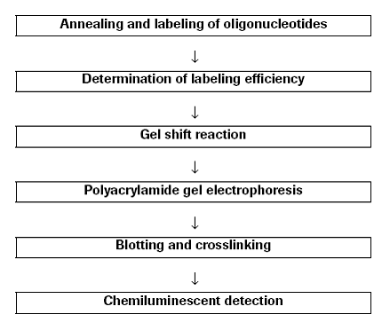
Procedures |
|
Annealing and labeling of oligonucleotides |
1.
2.
3.
4.
5.
6.
7.
8. | Mix solutions of complementary oligonucleotides in TEN-buffer in a molar ratio of 1:1.
Incubate for 10 min at 95°C.
Cool slowly to 15-25°C.
Dilute with sterile TEN-buffer to 3-4 pmol/μl.
Add 3.85 pmol double strand oligonucleotide and sterile double distilled water to a final volume of 10 μl to a reaction vial.
For the control reaction, add 1 μl control oligonucleotide and 9 μl sterile double distilled water to a reaction vial.
Add the following on ice:
5x labeling buffer 4 μl
CoCl2-solution 4 μl
DIG-ddUTP solution 1 μl
Terminal transferase 1 μl
Mix and centrifuge briefly.
Incubate for 15 min at 37°C, then place on ice.
Stop the reaction by adding 2 μl 0.2 M EDTA (pH 8.0).
Add 3 μl double distilled water to a final volume of 25 μl, to obtain a final concentration of 4 ng/μl or 0.155 pmol/μl of the labeled oligonucleotide.
|
Determination of labeling efficiency |
1.
2.
3.
4.
5.
6.
7.
8.
9.
10. | Apply a 1 μl spot of tubes 1-5 from your labeled oligonucleotide and the labeled control to the nylon membrane.
Fix the nucleic acid to the membrane by cross linking with UV-light or baking for 30 min at 120℃.
Transfer the membrane into a plastic container with 20 ml of washing buffer. Incubate under shaking for 2 min at 15 to 25°C.
Incubate in 10 ml of blocking solution for 30 min.
Incubate in 10 ml of antibody solution for 30 min.
Wash with 10 ml of washing buffer, 2 x 15 min.
Equilibrate 2-5 min in 10 ml of detection buffer.
Place membrane with DNA side facing up on a development folder (or hybridization bag) and apply 0.1 ml of CSPD® working solution.
Immediately cover the membrane with the second sheet of the folder to spread the substrate evenly and without air bubbles over the membrane.
Incubate for 5 min at 15 to 25°C. Squeeze out excess liquid and seal the edges of the development folder.
Note: drying of the membrane during exposure will result in dark background.
Incubate the damp membrane for 10 min at 37°C to enhance the CSPD chemiluminescent reaction.
Expose to X-ray film or imaging device.
Note: luminescence continues for at least 48 hours. The signal increases in the first few hours after initiation of the detection reaction until it reaches a plateau where signal intensity remains almost constant during the next 24-48 hours.
Multiple exposures can be taken to achieve the desired signal strength.
|
Gel shift reaction |
1.
2.
3.
4.
5.
| Mix on ice:
Binding buffer
DIG-labeled oligonucleotide
Protein
Double distilled water
Mix carefully and incubate for 15-30 min at 15-25℃.
Place tube on ice.
Add to each sample 5 μl of loading buffer with bromophenol blue.
Apply samples immediately to a pre-electrophoresed polyacrylamide gel.
|
Polyacrylamide gel electrophoresis |
1.
2.
3.
4.
5.
| One day before
Prepare a native polyacrylamide gel of 4% or 6% acrylamide in 0.5 x TBE buffer.
Note: prepare the gel the day before use to make sure that the gel is completely polymerized.
The gel must be pre-run.
Load the samples to the gel.
Note: before loading samples, clean sample wells to remove APS, urea and residual polyacrylamide to ensure sample application without diffusion.
Run a 10 cm x 10 cm x 0.1 cm PAGE at 80 V.
Note: for other gel sizes use 8 V/cm.
Run dye 2/3 of the way to the bottom of the plates.
|
Blotting and crosslinking |
1.
2.
3.
4.
5.
6.
7.
8.
9.
| After electrophoresis: remove one glass plate carefully from the gel.
Equilibrate a sheet of nylon membrane trimmed to the size of the gel for 5 min in transfer buffer (0.5x TBE buffer).
Place equilibrated nylon membrane carefully onto the gel.
Note: avoid air bubbles between gel and filter.
Place 4 layers of gel-sized Whatman 3MM papers, presoaked in transfer buffer on the filter.
Roll with a glass rod or a pipette over the 4 layers Whatman 3 MM paper to remove air bubbles.
Remove pad of Whatman 3MM paper/nylon membrane/gel from the other glass plate.
Add 4 layers of pre-soaked Whatman 3MM papers on the other side of the gel.
Place resulting sandwich between the electrodes of the electroblotting device.
Perform transfer for a 10 cm x 10 cm x 0.1 cm PAGE for 30 min at 400 mA (NOVEX System: 60 min; 30 V; 300 mA).
|
Crosslinking of oligonucleotides |
| Bake at 120°C: bake the membrane for 15-30 min
Or:
Place the membrane on a Whatman 3 MM paper presoaked with 2x SSC, cross-link at 120 mJ in e.g. a stratalinker or with a transilluminator for e.g. 3 min which has to be tested empirically.
|
Chemiluminescent detection |
1.
2.
3.
4.
5.
6.
7.
8.
9.
| Rinse membrane briefly (1-5) min in washing buffer.
Incubate for 30 min in 100 ml of blocking solution.
Incubate for 30 min in 20 ml of antibody solution.
Wash 2 x 15 min in 100 ml of washing buffer.
Equilibrate 2-5 min in 20 ml of detection buffer.
Place membrane with DNA side facing up on a development folder (or hybridization bag) and apply 1 ml CSPD working solution.
Immediately cover the membrane with the second sheet of the folder to spread the substrate evenly and without air bubbles over the membrane.
Incubate for 5 min at 15 to 25°C.
Squeeze out excess liquid and seal the edges of the development folder.
Note: Drying of the membrane during exposure will result in dark background.
Incubate the damp membrane for 10 min at 37°C to enhance the luminescent reaction.
Expose to X-ray film for 15-25 min or imaging device at 15–25°C.
Note: luminescence continues for at least 48 hours. The signal increases in the first few hours after initiation of the detection reaction until it reaches a plateau where signal intensity remains almost constant during the next 24-48 hours.
Multiple exposures can be taken to achieve the desired signal strength.
|
Materials |
| Washing buffer:0.1 M maleic acid, 0.15 M NaCl, pH 7.5 (20°C); 0.3% (v/v) Tween-20, 15 to 25°C, stable.
Maleic acid buffer:0.1 M maleic acid, 0.15 M NaCl, adjust with NaOH (solid) to pH 7.5 (20°C), 15 to 25°C, stable.
Detection buffer:0.1 M Tris-HCl, 0.1 M NaCl, pH 9.5 (20°C), 15 to 25°C, stable.
TEN-buffer:10 mM Tris, 1 mM EDTA, 0.1 M NaCl, pH 8.0, 15 to 25°C, stable.
Blocking stock solution:Dissolve blocking reagent 10% (w/v) in maleic acid buffer under constantly stirring on a heating block (65°C) or heat in a microwave oven, autoclave. The solution remains opaque. Stable for 4 weeks at 2 to 8°C if kept sterile.
1x Blocking solution:Prepare a 1x working solution by diluting the 10x blocking solution 1:10 in maleic acid buffer. Always prepare fresh.
Antibody solution:Centrifuge Anti-Digoxigenin-AP for 5 min at 10,000 rpm in the original vial prior to each use, and pipet the necessary amount carefully from the surface. Dilute Anti-Digoxigenin-AP 1:10 000 (75 mU/ml) in blocking solution. And 12 h at 2 to 8°C binding to the DIG-labeled probe.
CSPD working solution:Dilute 0.1 mg/ml stock solution 1:100 in detection buffer, and 2 to 8°C protected from light chemiluminescent detection.
|